


A fungal mycelium in three dimensions
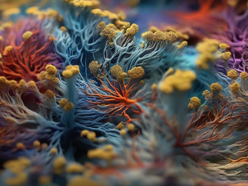
Three-dimensional hyphal growth: what would that look like?

Everybody has to have a dream. In my case, I have several science related dreams, and one of them is to see one of our lab’s saprobic fungi in actual three dimensions (3-D). I mean one fungus from our set of about 30 isolates with which we have worked during the past 10 years or so. We know a lot about these fungi, but ironically not what they really look like. I’d also be fine with any other fungus. I would love to see what a fungus looks like in 3-D for just about any fungus. So much so that every time I teach my fungal biology course I mention this challenge, and offer a cash prize to anybody who has an idea that will let us do this.
What do I mean? And why is this interesting and relevant, you ask?
Well, fungi are typically cultured on Petri dishes. That is because they are also regarded as microbes (which they are), and thus the same method for growing bacteria has been adopted for these organisms as well. Makes sense in a way. However, a Petri dish is pretty much a 2-D-system. And that is the problem: because fungi are also operating at a larger scale than just microscopic, and this unfolds actually in 3-D. This means, the fungus will basically expand from a germinating spore in all directions. And given that the hallmark of fungi is invasive growth, they grow their mycelia inside of their substrate, not on top of a thin layer of agar in a Petri dish. So this is the challenge: since the fungus grows in 3 dimensions inside of a solid substrate, we do not know what this looks like, since we cannot see it or visualize it. We cannot look inside of a substrate that has a fungus growing internally.
This means that everything we see in a Petri dish has to be an artifact to a certain degree. All these beautiful structures and colors, they do not reflect reality. But this is just a 2-D representation of fungal growth that in reality unfolds in 3 dimensions inside of a solid substrate.
What does this reality look like? Nobody really knows. Now, I have relatively small dreams, so I don’t need to see the 3-D structure of a fungus in its natural environment, like the soil. That would be incredibly difficult to impossible with current methods. My small dream is to see it in some sort of translucent medium, under lab conditions. And it also doesn’t need to be huge. Just big enough so that we can see it; I am thinking even smaller than a Petri dish would suffice. This would be enough to see growth unfold in three dimensions and observe it even with the naked eye or under a dissection scope.
My gut feeling is that this could reveal some interesting new traits that are currently completely non-imagined and untapped. Perhaps fungi explore dimensions to a different degree, so perhaps they have a certain shape in 3-D? Or do they really all just make spheres, more or less? Maybe mycelia of different species interacting in 3-D could reveal some interesting patterns; perhaps they sort themselves in ways that avoids some more direct interaction?
Got any ideas how to do this? Please let me know! It will need to be a translucent medium inside of which a saprobic fungus could grow (so there need to be nutrients and carbon, and ideally aeration).
Could the medium be made thicker and transparent? Like maybe in a glass cube or jar? Growth could be visualized that way. Then it could be digitized as suggested in Jon’s post.
Freeze it, then shave, take a picture, shave, take a picture. Repeat.
Then reassemble digitally and you can fly through it all. They did this with the human body.